Case 199
Case History
A 58-year-old woman presents with a palpable left breast lump.
Physical Examination
• left breast: 1 cm soft nodule in the upper outer quadrant
• right breast: normal exam
Mammogram
Mass
• architectural distortion (Fig. 199–1)
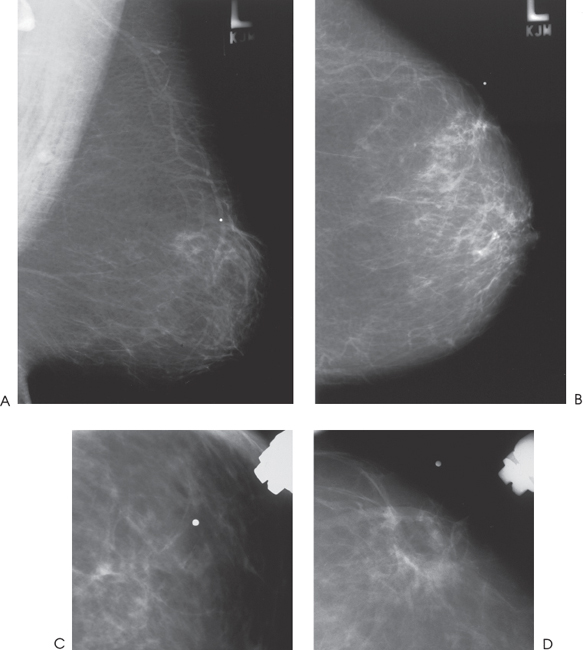
Figure 199–1. Left breast mammograms: Although the breast is predominantly fatty in composition, no mass is associated with the radiographic marker. The marker identifies the location of a palpable lump. In the CC spot compression view, there is a subtle small area of architectural distortion associated with the radiographic marker. (A). Left MLO mammogram. (B). Left CC mammogram. (C). Left MLO spot compression mammogram. (D). Left CC spot compression mammogram.
Ultrasound
Low Frequency
Frequency
• 7 MHz
Mass


Stay updated, free articles. Join our Telegram channel

Full access? Get Clinical Tree


