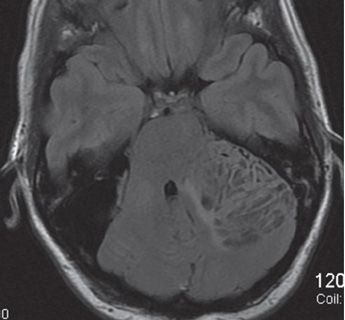
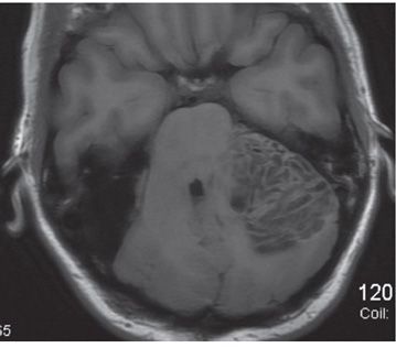
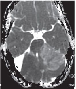
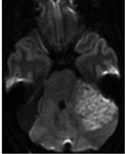

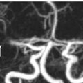
FINDINGS Figure 199-1. Axial T2WI through the posterior fossa. There is a well-circumscribed 5.4 cm × 3.8 cm exophytic left cerebellar mass insinuating itself into the left cerebellopontine angle (CPA) (arrows), compressing the left brachium pontis medially and mildly rotating the brainstem. The fourth ventricle is compressed. Mass is composed of a lacy and striped pattern of alternating isointensity and hyperintensity. There is a large extraaxial cyst on the right (star) between the cerebellum and the petrosal surface consistent with a cyst. Supratentorially (not shown) there are multiple flame-shaped subcortical T2 hyperintensities radiating and tapering unto the ventricular walls consistent with focal cortical dysplasia (FCD). Figures 199-2 and 199-3. Axial FLAIR and T1WI through the mass. There are alternating hypoisointense and isointense stripes through the mass. The hypointense areas correspond to the hyperintensities on T2WI consistent with cystic areas, while the isointense areas are the hypertrophied cerebellar folia. Figures 199-4 and 199-5


Stay updated, free articles. Join our Telegram channel

Full access? Get Clinical Tree


