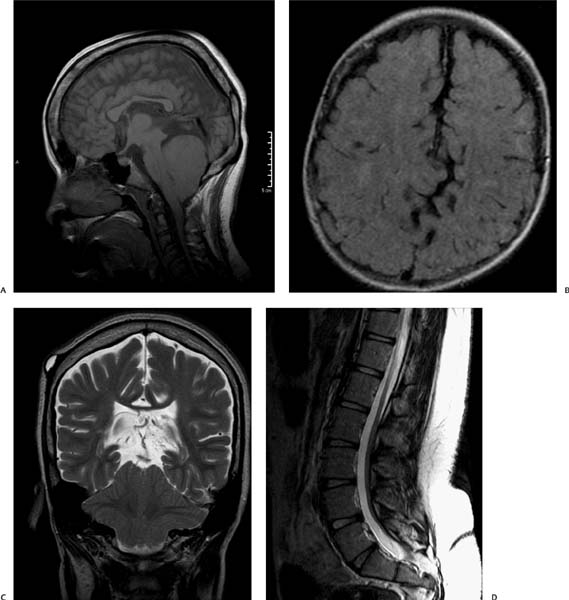
Case 2 A 25-year-old patient initially presenting with lower extremity paralysis, respiratory distress, and impaired swallowing. (A) Sagittal T1-weighted image (WI) of the brain shows inferior protrusion of the cerebellar vermis through a widened foramen magnum (asterisk). Note the tectal beaking (arrow) and dysgenesis of the corpus callosum (arrowhead). The 4th ventricle is effaced. (B) Axial fluid-attenuated inversion recovery image of the brain demonstrates an interdigitation of the sulci of the brain in the midline (arrows) secondary to hypoplasia of the cerebral falx. (C) Coronal T2WI of the brain shows a towering cerebellum and inferior tilting of the cerebellar tonsils through the foramen magnum (white arrows) along with the vermis (asterisk). (D) Sagittal T2WI of the spine shows posterior dysraphism of the sacrum (arrows) with an associated sinus tract (arrowhead).
Clinical Presentation
Imaging Findings
Differential Diagnosis
Stay updated, free articles. Join our Telegram channel

Full access? Get Clinical Tree


