 Clinical Presentation
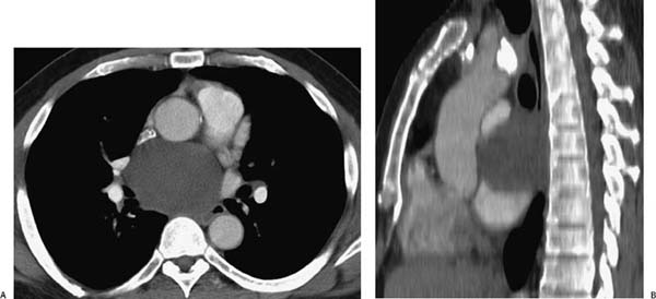
A 52-year-old man with cough and dysphagia and abnormal findings on chest radiograph.
 Imaging Findings
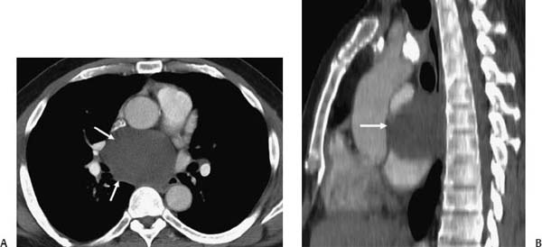
(A, B) Contrast-enhanced thoracic computed tomography (CT). Axial (A) and sagittal (B) re-formations demonstrate a low-density mass with a cystic appearance in the subcarinal region (arrows, A) that has a mild mass effect on the esophagus, which is seen between the aorta and the cystic mass (arrow, B).
 Differential Diagnosis
• Bronchogenic cyst (BC):
Stay updated, free articles. Join our Telegram channel

Full access? Get Clinical Tree


