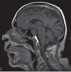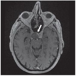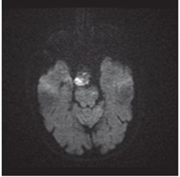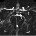


FINDINGS Figure 2-1. Axial T2WI through the sella turcica. There is a slightly hyperintense (to normal brain) mass, involving the sella/suprasellar region and right cavernous sinus (arrows). Figures 2-2 and 2-3. Axial and sagittal post-contrast T1WI, respectively. The mass is essentially nonenhancing and separate from the laterally displaced mildly enhancing pituitary gland (arrows). Figure 2-4. Axial DWI through the mass. The tumor is hyperintense which aids in the visual detection of the lesion and suggests hypercellularity.
DIFFERENTIAL DIAGNOSIS Meningioma, macroadenoma, metastasis, chordoma.
DIAGNOSIS Prostate cancer metastasis.
DISCUSSION
Stay updated, free articles. Join our Telegram channel

Full access? Get Clinical Tree


