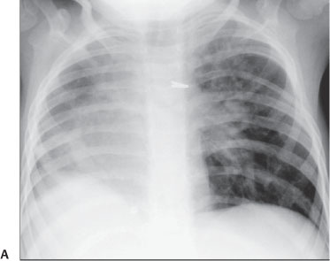
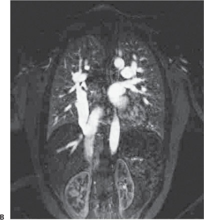
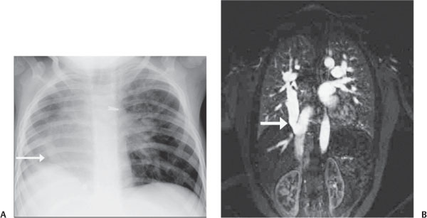
Case 2 A 2-year-old boy with chronic cough. (A) Anteroposterior chest radiograph. There is a small right lung with shift of the heart and mediastinum to the right. Additionally, there is a curved opacity near the elevated right hemidiaphragm (arrow). (B) Coronal magnetic resonance (MR) image demonstrates an anomalous right pulmonary venous connection to the inferior vena cava (arrow). • Scimitar syndrome:
Clinical Presentation
Further Work-up
Imaging Findings
Differential Diagnosis
![]()
Stay updated, free articles. Join our Telegram channel

Full access? Get Clinical Tree


