Case 2

 Clinical Presentation
A 24-year-old woman with a history of recurrent urinary tract infection since childhood.
 Imaging Findings

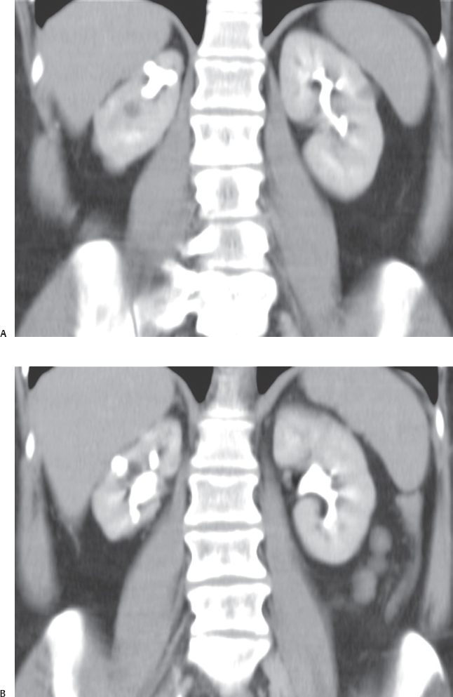
(A) Thick-slab multiplanar reconstruction (MPR) image from the excretory phase of a computed tomographic (CT) intravenous pyelogram (IVP) study shows that the right kidney is small. The outline is irregular because of asymmetric parenchymal loss caused by scarring (arrows) overlying dilated upper pole calices (arrowheads). The left kidney is normal. (B) Thick-slab MPR image anterior to the level of Figure A shows scarring (arrows) overlying dilated interpolar and lower pole calices (arrowheads). There is no dilatation of the right renal pelvis (asterisk). The left kidney is normal.
 Differential Diagnosis
• Reflux nephropathy:
Stay updated, free articles. Join our Telegram channel

Full access? Get Clinical Tree


