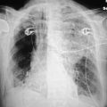
A team approach is needed to obtain a portable chest radiograph, and the team should include technologists, radiologists, and ICU personnel. There should be close cooperation between ICU nurses, respiratory therapists, and other ICU personnel in assisting the technologist while positioning an unstable patient. They should help remove unnecessary and unwanted tubes and lines from the patient’s chest before taking the film because these can be misinterpreted and thought to be in the heart or lungs. Furthermore, every attempt must be made to obtain the film with the patient upright and at the end of deep inspiration. If the radiograph is obtained in expiration when the patient is supine, the findings can be misleading. Finally, it is imperative that there be rapid communication between the radiologist and the ICU team about the findings seen on a chest radiograph.4
Because the quality of the image often determines the diagnostic information that can be obtained from the radiograph, it is necessary to review the factors affecting the film, such as contrast, noise, and spatial resolution.
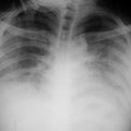
Contrast represents the variation of film density between one part of the film and another. Ideally one should obtain a film with the highest contrast throughout, and in the chest x-ray this is generally measured from the darkest area (Dmax), which is the lung, to the lightest, white area (Dmin), which is bone. Contrast is the result of the differences between tissues as an x-ray photon passes through the object. The important parameters are thickness and density of the material through which the x-ray passes and the amount of absorption of the photon that occurs. In the diagnostic radiology range (60-150 KVp) the absorption coefficient has a direct relationship to the atomic number of the tissue through which the photon passes. In this range, the absorption coefficient is related to the cube of the atomic number. Objects with a high atomic number have a different contrast resolution than do objects with a lower atomic number. This permits us to differentiate structures because in the chest x-ray the heart has a different density, and a different atomic number, than the surrounding lung. The ribs and bony structures are made up principally of calcium, which has a higher atomic number than the lung and thus absorbs more of the x-ray beam, and are therefore easily visualized.5
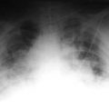
Noise degrades an image. It is generally seen as grainy mottle pattern on the x-ray. This is due to the statistical difference in the number of x-ray photons striking the film in different areas. In some areas, there are insufficient numbers of photons, so only a part of the film is exposed, producing a mottled appearance.
Spatial resolution is the ability of an x-ray to depict closely spaced objects. High spatial resolution produces sharper edges in an object than does low resolution. Image sharpness refers to how sharp the edge of an object is when seen adjacent to other objects.6
The key to optimizing the technical quality of a chest radiograph is to minimize variation in the technical parameters from day to day. One should instruct the technologist to choose the same patient position each day and to keep a written record of the previous successful techniques at each patient’s bedside. The first time a film is obtained on an ICU patient, the technologist should show that film to the ICU radiologist, who will determine if the technique is adequate. If so, this technique should subsequently be used on all follow-up films so that the exposure is similar and changes in the most recent x-ray are due to changes in the patient’s condition, rather than changes induced by differences in radiologic technique.7
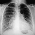
It is necessary to keep in mind that portable chest films are performed in the anterior posterior (AP) position, resulting in a 15–20% magnification factor to the cardiac shadow. One should be cautious when making the diagnosis of cardiomegaly on an AP film. Remember that one needs an adequate inspiration (9–10 ribs) to establish that diagnosis.8
Exposure variations constitute one of the main difficulties in portable radiography. Photo-timing devices, used in the main department, are generally not used for bedside films.9 Generally, the
exposure must be estimated by the technologist. The principal variables that affect quality film include the kilovolt potential (KVp) and milliampere second (mAs) used, the patient’s size, the use of a grid, the film system speed, and the distance between the x-ray tube and the image receptor.
While it is possible to vary the kVp setting, this is normally not done because altering the kVp even minimally will produce large changes in exposure. On a portable chest film, a 20% change in kVp will be the equivalent of doubling the mAs.
The mAs is the product of tube current in milliamperes (mA) and exposure time in seconds (s). Normally the mA is fixed, and change occurs due to variations made in the time of exposure. Generally, the technologist adjusts the mAs to achieve correct film exposure.
Thus normally the kVp is kept constant and the mAs is varied. If the distance between tube and film is increased, the mAs needs to be increased. Increasing the mAs poses a problem especially in large patients or those who are unable to hold their breath because lack of sharpness occurs on exposure times greater than 10 ms.
Patient size is another variable, but an experienced technologist can quickly estimate the need for increased exposure when needed. The degree of pathology in the lung also contributes to the necessity for increasing exposure because abnormalities that increase density in the lung cause more of the x-ray beam to be absorbed.
In portable radiography, the distance between the x-ray tube and the film is not fixed. It is extremely important to keep tube–film distance unchanged from day to day. Exposure is inversely related to distance, so small variations in distance can account for large changes in exposure. A change of 6 in. from one day to the next can result in a 50% variance in film exposure; 50 in. is the optimal distance from tube to film.
Related posts:


Stay updated, free articles. Join our Telegram channel

Full access? Get Clinical Tree


