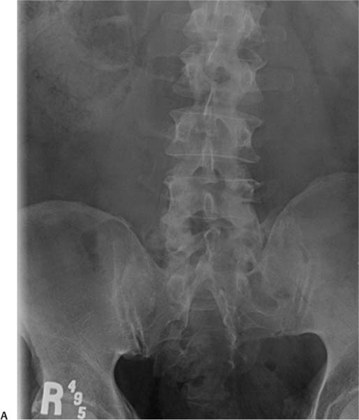
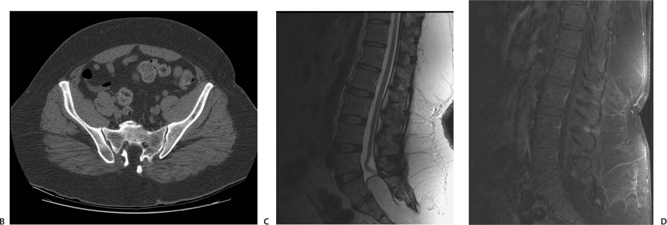
Case 20 A patient with urinary incontinence and a subcutaneous mass in the sacral region. (A) Anteroposterior radiograph of the lumbar spine shows dysraphism of the sacrum (arrows). (B) Axial computed tomography (CT) scan of the pelvis without contrast demonstrates the lack of fusion of the posterior elements of the sacrum (black arrows). Fat corresponding to the lipoma occupies the defect (white arrow). (C) Sagittal T2-weighted image (WI) of the lumbar spine shows the conus descending to L4 (white arrow). The lipoma extends into the spinal canal (asterisk). The black arrow points to the placode-lipoma interface. (D) Sagittal T1WI of the lumbar spine with fat saturation and contrast; note the fat suppression of the lipoma (asterisk).
Clinical Presentation
Further Work-up
Imaging Findings

Stay updated, free articles. Join our Telegram channel

Full access? Get Clinical Tree


