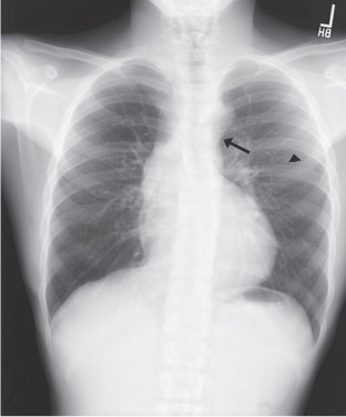
Case 20 A hypertensive teenager with lower blood pressures in the lower extremities than in the upper extremities. Frontal chest radiograph demonstrates ectasia of the ascending aorta and an enlarged aortic knob with a “notch” between the aortic knob and the proximal descending thoracic aorta (arrow). There is also mild rib notching, best seen in the posterior aspect of the left sixth rib (arrowhead). • Coarctation of the aorta: The findings of a prominent ascending aorta, enlarged aortic knob, notch between the aortic knob and proximal descending thoracic aorta, and rib notching are highly suggestive of coarctation of the aorta. • Aortic stenosis:
Clinical Presentation
Imaging Findings
Differential Diagnosis
![]()
Stay updated, free articles. Join our Telegram channel

Full access? Get Clinical Tree


