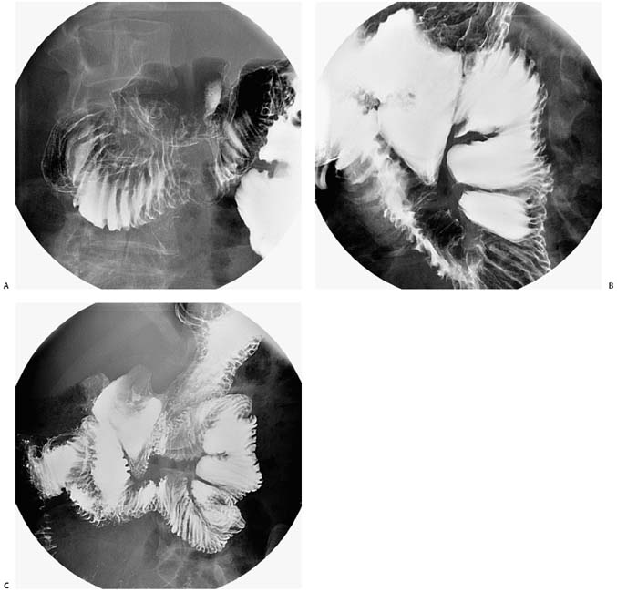
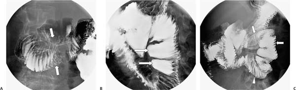
Case 20 A 39-year-old woman presents with nausea and abdominal distension. (A) Upper gastrointestinal (GI) barium study shows stacks of straight duodenal folds and duodenal dilatation (arrows). (B) The proximal jejunum is dilated with contrast-filled pseudosacculations (arrows). (C) Delayed image shows dilatation of the duodenum and jejunum (arrows) with stacks of thin, straight folds throughout the jejunum (arrowhead). • Scleroderma: This is the most likely diagnosis, given the stacks of thin, straight duodenal and jejunal folds (hidebound). Note the dilated small bowel, which is most consistent with scleroderma. • Small-bowel obstruction: This may present with dilated loops, but without stacked small-bowel folds. • Pseudo-obstruction:
 Clinical Presentation
 Imaging Findings
 Differential Diagnosis
![]()
Stay updated, free articles. Join our Telegram channel

Full access? Get Clinical Tree


