Case 201
Case History
A 45-year-old woman presents with thickening in the right breast, which she thinks might be a pulled muscle.
Physical Examination
• right breast: large, mildly tender firm area extending from 9:00 to 12:00 in the right breast
• left breast: normal exam
Mammogram
Mass (Fig. 201–1)
• margin: ill defined
• shape: oval
• density: isodense
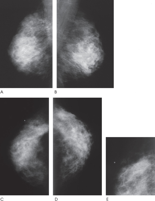
Figure 201–1. There is an ill-defined mass in the right upper outer quadrant. The size of the mass is difficult to determine because of the surrounding heterogeneous parenchymal density. (A). Right MLO mammogram. (B). Left MLO mammogram. (C). Right CC mammogram. (D). Left CC mammogram. (E). Right MLO spot compression mammogram.
Ultrasound


Stay updated, free articles. Join our Telegram channel

Full access? Get Clinical Tree


