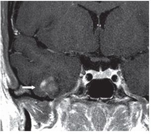
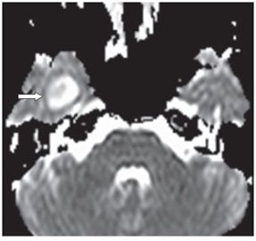
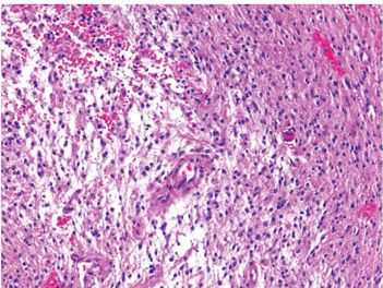
FINDINGS Figure 202-1. Axial FLAIR through the inferior temporal lobes. There is a well-circumscribed hyperintense right temporal lobe mass (arrow). There is no edema. Figure 202-2. Coronal post-contrast T1WI through the mass. There is mildly heterogeneous solid enhancement (arrow). Figure 202-3. Axial ADC map through the mass. There is high diffusion with a central normal focus (arrow). Figure 202-4. Photomicrograph shows compact cellular (upper right) and loose myxoid (lower left) areas. Cells are elongated in compact areas (H&E stain).
DIFFERENTIAL DIAGNOSIS Stroke, abscess, demyelination, pilocytic astrocytoma (PA), ganglioglioma, pleomorphic xanthoastrocytoma (PXA), dysembryoplastic neuroepithelial tumor (DNET), oligodendroglioma.
DIAGNOSIS Pilocytic astrocytoma (PA) lobar.
DISCUSSION


Stay updated, free articles. Join our Telegram channel

Full access? Get Clinical Tree


