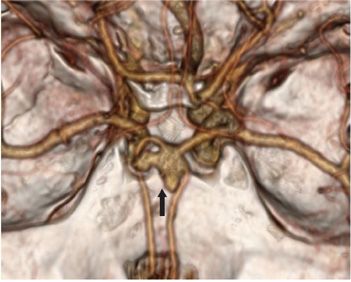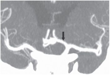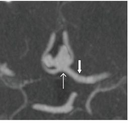


FINDINGS Figure 203-1. Axial NCCT of the brain through the supra sella cistern. There is a 1-cm pear-shaped hyperdense structure in the interhemispheric fissure separating the posterior aspect of the bilateral gyrus rectus (arrow). This structure is about the same size as it was 8 years earlier when it was first noticed on NCCT. It was suggested that it could represent an aneurysm. There was no hemorrhage on the previous or on the present CT. Figure 203-2. 3D volume-rendered CTA submentovertical (SMV) view. There is a 1.2-cm lobulated outpouch consistent with an aneurysm (arrow) of the anterior communicating artery emanating from the junction of the left A1, and bilateral A2s and absence of right A1 segment. Aneurysm is pointing superiorly and anteriorly separating the two A2s. It has a clearly defined neck that measures about 4.5 mm. There is a right-sided lobule or daughter aneurysm pointing to the right. The other vascular structures appear normal with no evidence of spasm. Figures 203-3 and 203-4. Coronal and axial MIP CTA, respectively. The relationship of the aneurysm neck (line arrow) to the other vascular structures is defined. The right A1 is missing presumably congenital, a normal variant. Normal left A1 (arrow).
Differential Diagnosis N/A.
DIAGNOSIS
Stay updated, free articles. Join our Telegram channel

Full access? Get Clinical Tree


