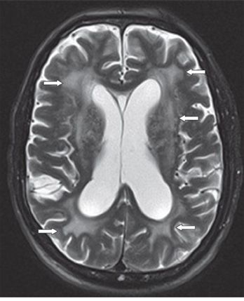
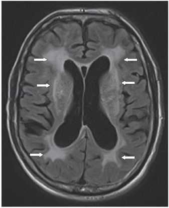
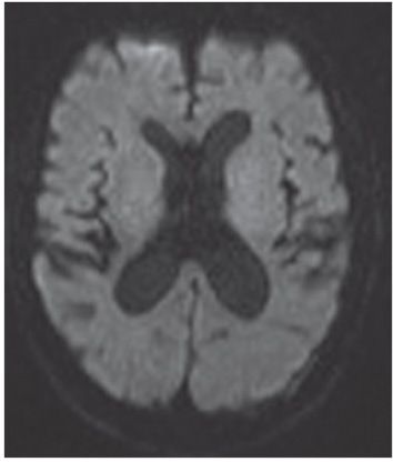
FINDINGS Figure 204-1. Axial NCCT through the corona radiata. There are patchy periventricular white matter (WM) hypodensities (arrows) and a mild degree of global brain volume loss. Figures 204-2 and 204-3. Axial T2WI and FLAIR respectively through the corona radiata. There is mild to moderate volume loss. There are both patchy and confluent WM hyperintensities in bilateral corona radiata (arrows). Figure 204-4. Corresponding axial DWI shows no acute infarctions.
DIFFERENTIAL DIAGNOSIS Leukoaraiosis, cerebral atherosclerosis, amyloid angiopathy, transependymal cerebrospinal fluid (CSF) flow, radiation-induced damage, cerebral autosomal dominant arteriopathy with subcortical infarcts and leukoencephalopathy (CADASIL).
DIAGNOSIS Leukoaraiosis.
DISCUSSION


Stay updated, free articles. Join our Telegram channel

Full access? Get Clinical Tree


