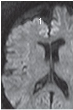
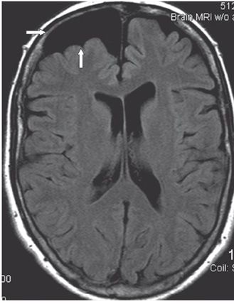
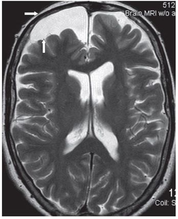
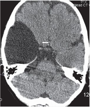
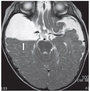
FINDINGS Figure 205-1 Axial post-contrast CT. There is a right frontal extraaxial cerebrospinal fluid (CSF) density collection with compression of the right frontal lobe (vertical arrow). There is mild thinning and remodeling of the overlying right frontal bone (transverse arrow). Figures 205-2 to 205-4. Axial DWI, FLAIR, and T2WI, respectively, through the lesion. The collection follows CSF intensity on all sequences. There is compression of the frontal lobe (vertical arrows). Thinning of the right frontal bone is again demonstrated (transverse arrows). Figures 205-5 and 205-6. Axial NCCT and axial T2W MRI, respectively, through the right middle cranial fossa in a 3-year-old boy. These demonstrate an arachnoid cyst (AC) in a more typical location in the right middle cranial fossa. There is smooth compression of the frontal and temporal lobes (arrows) with overlying right temporal bone remodeling and thinning seen on the CT. In these two cases like in all cases, the mass is extraaxial to cortical gray matter (GM).
DIFFERENTIAL DIAGNOSIS AC, epidermoid cyst, neurocysticercosis, porencephalic cyst (PC), pilocytic astrocytoma, hemangioblastoma.
DIAGNOSIS Arachnoid cyst (AC).
Stay updated, free articles. Join our Telegram channel

Full access? Get Clinical Tree


