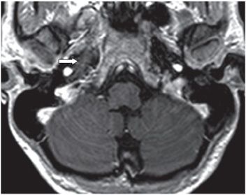
FINDINGS Figure 206-1. Axial T2WI through the petrous apices. There is hyperintensity of the right petrous apex (arrow). Figure 206-2. Axial post-contrast T1WI. There is subtle enhancement of the bone and surrounding soft tissues (arrow).
DIFFERENTIAL DIAGNOSIS Trapped fluid, petrous apicitis, cholesterol granuloma, cholesteatoma, tumor.
DIAGNOSIS Petrous apicitis.
DISCUSSION CT findings of petrous apicitis include loss of aeration or trabecular pattern with hypodensity of the petrous apex with variable contrast enhancement. The MR T1WI often shows a hypointense petrous apex with contrast enhancement. Lesion is hyperintense on FLAIR and T2WI and may show some expansion of the petrous apex.
Stay updated, free articles. Join our Telegram channel

Full access? Get Clinical Tree


