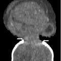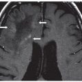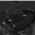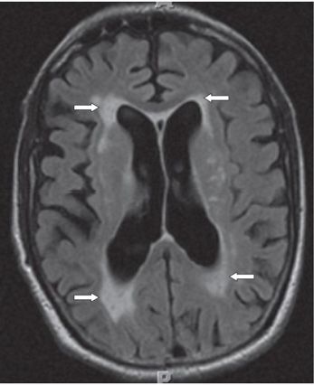
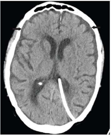
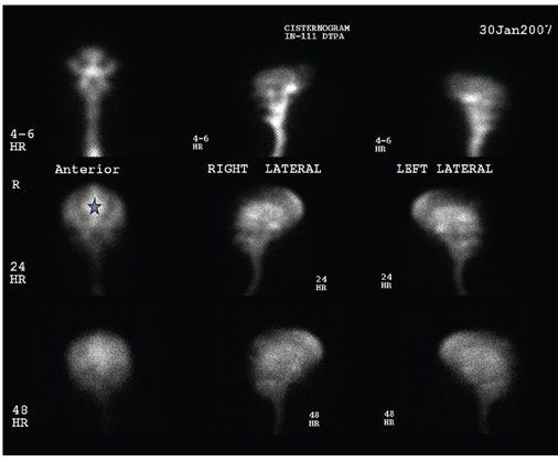
FINDINGS Figure 209-1. Axial T2WI through lateral ventricles. There is ventricular enlargement out of proportion to sulcal dilation. There is bilateral frontal and occipital periventricular caps (arrows). Figure 209-2. Axial FLAIR through the body of the lateral ventricle. There is confluent periventricular hyperintensity over the frontal and occipital regions suggestive of transependymal cerebrospinal fluid (CSF) flow (arrows). Figure 209-3. Axial NCCT through the lateral ventricles following ventriculoperitoneal (VP) shunting. There is normalized caliber of the ventricles with resolution of periventricular caps and interval appearance of thin subdural collections (arrows). Figure 209-4. Pre-operative In-111 diethylene triamine pentaacetic acid (DTPA) cisternography. There is persistent radiotracer uptake in the lateral ventricles (star) at 24 hours.
DIFFERENTIAL DIAGNOSIS Normal aging brain, Alzheimer disease, vascular dementia, sporadic subcortical arteriosclerotic encephalopathy, Parkinson disease, normal pressure hydrocephalus (NPH).
Stay updated, free articles. Join our Telegram channel

Full access? Get Clinical Tree


