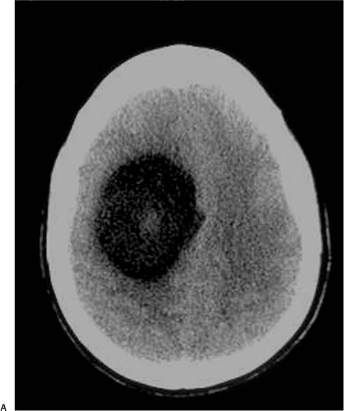
Case 21 A 25-year-old woman presenting with headaches and papilledema. (A) Computed tomography (CT) scan without contrast shows a round, hypoattenuated lesion in the right frontal lobe (arrows). The lesion is located in the subcortical white matter and demonstrates a coarse central calcification (arrowhead). (B) Axial T2-weighted image (WI) shows a hyperintense subcortical mass (arrows) in the right frontal lobe; no vasogenic edema is evident. An area of low signal is seen in the center of the mass (arrowhead), representing the calcification evident on the CT scan. (C) Coronal fluid-attenuated inversion recovery image shows a mass in the right frontal lobe with heterogeneous signal (arrows). There is effacement of the right frontal horn (arrowhead) and displacement of the midline to the left. (D) Coronal T1WI with contrast shows mild enhancement of the right frontal mass.
Clinical Presentation
Further Work-up
Imaging Findings
Differential Diagnosis
Stay updated, free articles. Join our Telegram channel

Full access? Get Clinical Tree


