 Clinical Presentation
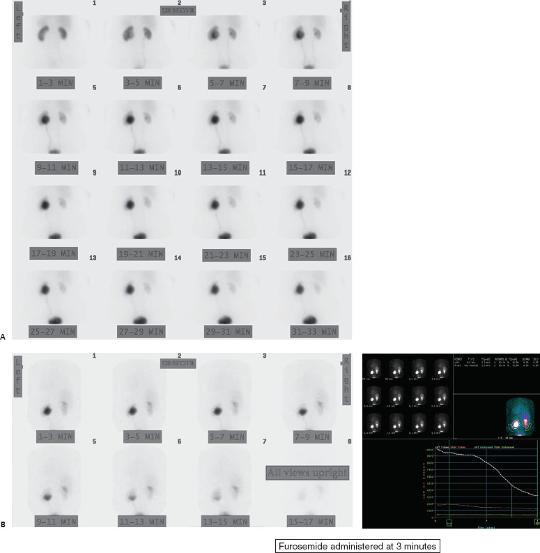
A 23-year-old woman with hydronephrosis detected on abdominal US.

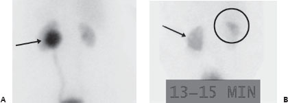
(A) Tc99m MAG3 renal scintigraphy demonstrates nearly symmetric renal parenchymal function (similar parenchymal counts at 1–3 minutes) but progressive radiotracer accumulation into a dilated renal pelvis on the left (arrow). Both ureters are seen and do not appear significantly dilated. (B) Following furosemide, there is rapid washout from the left collecting system (arrow) with a half-time of 9 minutes. Progressive physiologic excretion is seen in the gallbladder (circle), which can be seen with MAG3 (but not DTPA).
 Differential Diagnosis
• Dilated but nonobstructed upper renal collecting system: Renal pelvocaliceal dilatation but with rapid washout into the bladder after diuretic makes this the most likely diagnosis.
• Chronic reflux: This can also have renal pelvic dilatation and rapid washout. However, some degree of ureteral dilatation can usually be appreciated as well.
• Current renal obstruction: This will also have pelvocaliceal dilatation but will demonstrate poor washout after diuretic.
 Essential Facts


Stay updated, free articles. Join our Telegram channel

Full access? Get Clinical Tree


