Courtesy of K. Salzman, MD.

Courtesy of K. Salzman, MD.

Courtesy of K. Salzman, MD.

Courtesy of K. Salzman, MD.
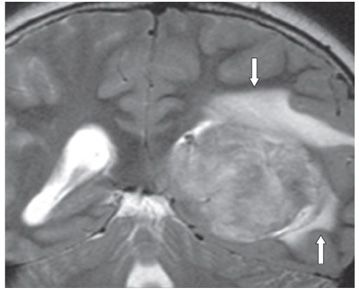
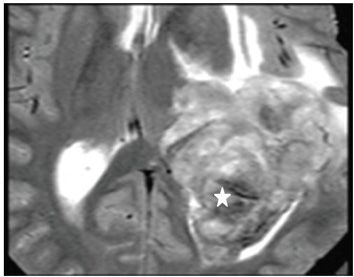
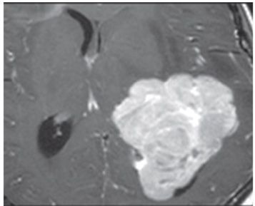
FINDINGS Figure 21-1. Coronal non-contrast T1WI through the trigone/occipital horns. There is a large isointense mass within the left trigone and occipital horn (star). There is blurring of the superior and lateral margins of the left trigone (arrows). Figure 21-2. Coronal T2WI through same level as in Figure 21-1. The left lateral ventricular mass is heterogeneously hyperintense with surrounding parenchymal vasogenic edema superiorly and laterally (arrows). Figure 21-3. Axial T2WI. There are hypointense areas within the posterior aspect of the mass (star) suggesting either calcifications or blood products. There is surrounding vasogenic edema anteriorly. There is no clear separation between the tumor and the anterior and lateral walls of the trigone suggestive of infiltration. Figure 21-4. Axial post-contrast T1WI. The mass is avidly contrast enhancing and lobulated with clearly defined margins. There is however possible infiltration of the anterolateral walls where the parenchymal vasogenic edema exists. There is local mass effect.
Stay updated, free articles. Join our Telegram channel

Full access? Get Clinical Tree


