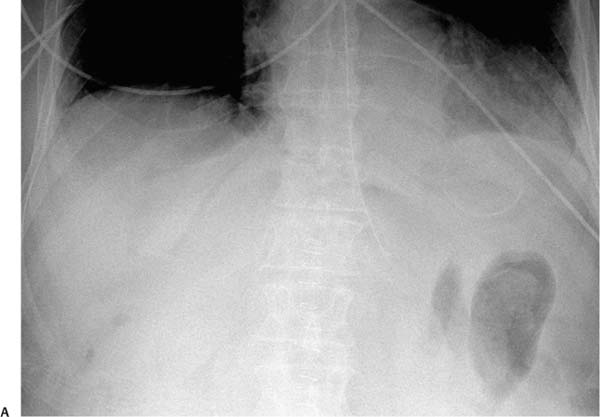
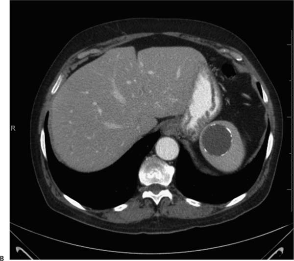
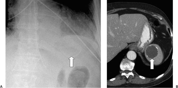
Case 21 A 70-year-old man presents with abdominal pain. (A) Abdominal radiograph shows a curvilinear calcification (arrow) projected over the left upper quadrant of the abdomen. (B) Infused abdominal computed tomography (CT) shows a round cystic lesion (arrow) with a calcified rim. • Simple splenic cyst: This is the most likely diagnosis for the CT finding of a fluid-density, nonenhancing, round structure in the spleen. The differential diagnosis for the curvilinear calcification seen on the radiograph is discussed in “Pearls & Pitfalls.” • Epidermoid cyst: This may have identical features, although it calcifies less commonly. • Echinococcal cyst:
 Clinical Presentation
Further Work-up
 Imaging Findings
 Differential Diagnosis
![]()
Stay updated, free articles. Join our Telegram channel

Full access? Get Clinical Tree


