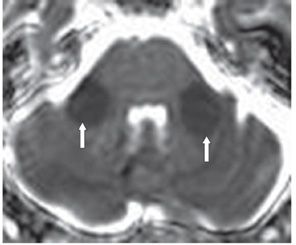
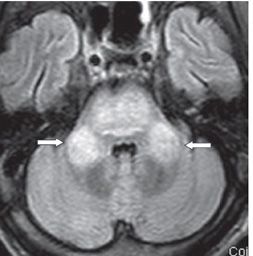
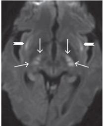
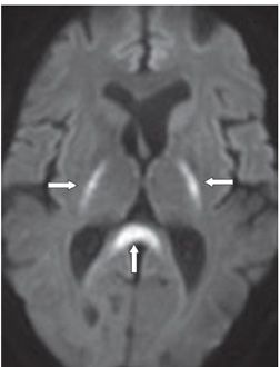
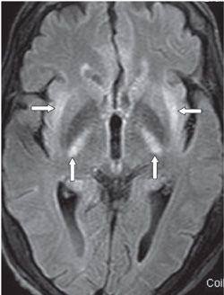

FINDINGS Figures 211-1 and 211-2. Axial MR DWI and corresponding ADC map through the brachium pontis. There is bilateral symmetrical ovoid area of diffusion restriction in the middle cerebellar peduncles (MCPs) (arrows). Figure 211-3. The corresponding axial FLAIR. There is hyperintensity not only in the bilateral MCP but also in the pons with a thin rim of normal pontine intensity (arrows). Figure 211-4. Axial DWI through the inferior basal ganglia. There is bilateral symmetrical restricted diffusion in the external capsules (chevrons), bilateral subthalamic regions (transverse arrows), and posterior limb of internal capsules (vertical arrows). Figures 211-5. Axial DWI and ADC map, respectively, through the basal ganglia. There is symmetrical restricted diffusion in the posterior limb of the internal capsules (transverse arrows) and in the splenium of the corpus callosum (vertical arrows). There is subtle hyperintensity in the head of caudate nucleus bilaterally. Figures 211-6 and 211-7


Stay updated, free articles. Join our Telegram channel

Full access? Get Clinical Tree


