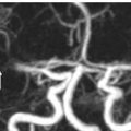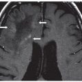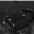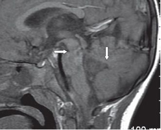
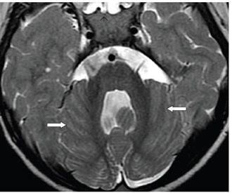
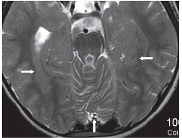
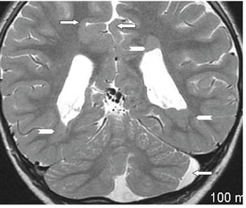
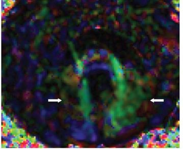
FINDINGS Figure 213-1. Axial NCCT through the upper cerebellum shows abnormal and irregular shape of the fourth ventricle with poor definition of the velum (posterior arrow). The interpeduncular cistern could be seen anteriorly (anterior arrow) suggesting a high-riding fourth ventricle. Figure 213-2. Sagittal post-contrast MRI T1WI shows widening of the fourth ventricle with the top of the fourth ventricle at the level of the interpeduncular cistern (transverse arrow). The superior velum is slightly flat (vertical arrow). Figure 213-3. Axial T2WI through the mid-fourth ventricle demonstrates widening of the fourth ventricle with a large nodule posteriorly. There is widening of the prepontine and cerebellopontine cisterns with a left pericerebellar cerebrospinal fluid (CSF) space widening. The cerebellar folia are obliquely directed and slightly asymmetric (arrows). Small but otherwise normal cerebellar peduncles and pons are present. Figure 213-4
Stay updated, free articles. Join our Telegram channel

Full access? Get Clinical Tree


