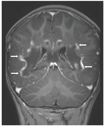
FINDINGS Figure 214-1. Axial T2WI through the trigones. There is bilateral symmetrical peritrigonal confluent white matter (WM) hyperintensity extending from the ventricular walls to the subcortical U fibers (stars) crossing the midline through the splenium of corpus callosum. Figure 214-2. Coronal post-contrast T1WI through the trigones. There is bilateral symmetrical centrally affected WM hypointense with peripheral wavy contrast enhancement following the pattern of the subcortical U fibers (arrows).
DIFFERENTIAL DIAGNOSIS Krabbe disease, metachromatic leukodystrophy (MLD), Alexander disease, Adrenoleukodystrophy (ALD), Canavan disease.
DIAGNOSIS Adrenoleukodystrophy (ALD).
DISCUSSION


Stay updated, free articles. Join our Telegram channel

Full access? Get Clinical Tree


