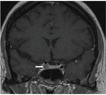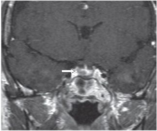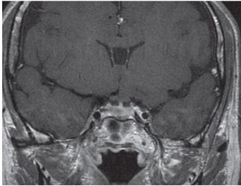


FINDINGS Figure 215-1. Coronal dynamic post-contrast T1WI. There is a 6-mm nonenhancing mass (arrow) in the right side of the pituitary gland. Figure 215-2. Delayed static high-resolution corresponding post-contrast T1WI in the same patient also shows the adenoma. Figure 215-3. Coronal 90-second dynamic post-contrast coronal T1WI in a companion case shows a right-sided adenoma (arrow). Figure 215-4. Coronal post-contrast delayed T1WI. The tumor is less conspicuous due to enhancement that makes it more difficult to differentiate from the adjacent gland. This case shows the advantage of using dynamic imaging for evaluation of microadenomas.
DIFFERENTIAL DIAGNOSIS Pituitary microadenoma, Rathke cleft cyst, pars intermedia cyst.
DIAGNOSIS Pituitary microadenoma.
DISCUSSION
Stay updated, free articles. Join our Telegram channel

Full access? Get Clinical Tree


