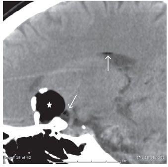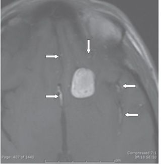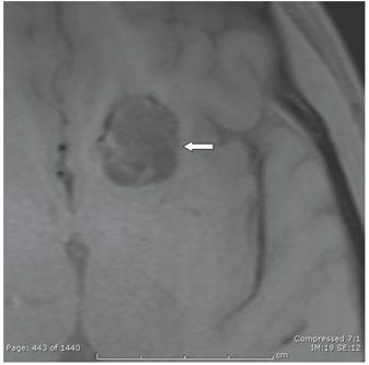


FINDINGS Figures 217-1 and 217-2. Coronal and sagittal reformatted NCCT through the planum, respectively. There is a fat density (star), extraaxial left supraclinoid mass with rim calcifications. In addition, there are scattered fat droplets throughout the subarachnoid spaces (arrows). Figure 217-3. Axial T1WI through the mass. There is a hyperintense left supraclinoid lesion with hypointense peripheral signal. Multiple punctate similar hyperintensities are present in the subarachnoid spaces (arrows). Figure 217-4. Axial fat saturation T1WI through the mass. There is suppression of the hyperintense signal within the left mass. The scattered hyperintense foci within the subarachnoid spaces bilaterally on the T1WI suppress on the fat sat T1WI as well, confirming the fatty consistency.
DIFFERENTIAL DIAGNOSIS Dermoid cyst, epidermoid cyst, craniopharyngioma, teratoma, lipoma.
DIAGNOSIS Ruptured dermoid cyst.
DISCUSSION
Stay updated, free articles. Join our Telegram channel

Full access? Get Clinical Tree


