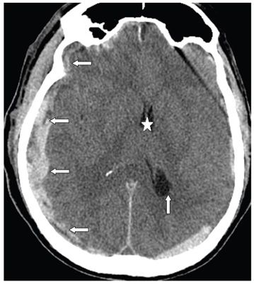
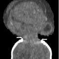
FINDINGS Figure 218-1. Axial NCCT through the level of the frontal horns at about 8 AM when she was brought into the emergency room. There are bilateral extraaxial crescentic collections larger and of mixed density (hypodense and hyperdense), on the left (arrows) and thinner iso- to hyperdense on the right (chevrons). There is a mild midline shift to the right (star). Figure 218-2. Axial NCCT through the same level as in Figure 218-1 about 3 hours later at about 11 AM when the patient’s condition deteriorated. There is now a larger mixed density (iso-hypo-hyperdense) crescentic extraaxial collection surrounding the right cerebral hemisphere (transverse arrows). The left extraaxial collection is now partially effaced. There is now a midline shift to the left (star). There is also dilatation (acute hydrocephalus) of the left trigone (vertical arrow) consistent with a trapped left lateral ventricle due to effacement and obstruction at the level of the foramen of Monro/third ventricle.
DIFFERENTIAL DIAGNOSIS Hyperacute subdural hematoma (HSDH), recurrent bleed into chronic subdural hematoma (CSDH).
DIAGNOSIS Hyperacute subdural hematoma (HSDH) with active bleed.
DISCUSSION


Stay updated, free articles. Join our Telegram channel

Full access? Get Clinical Tree


