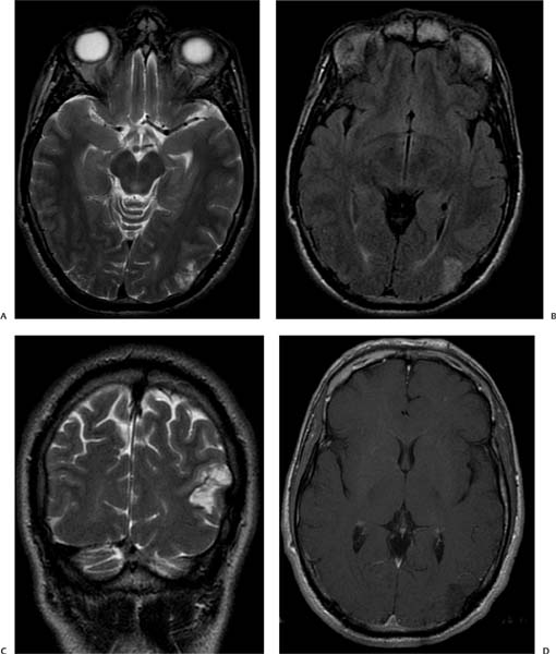
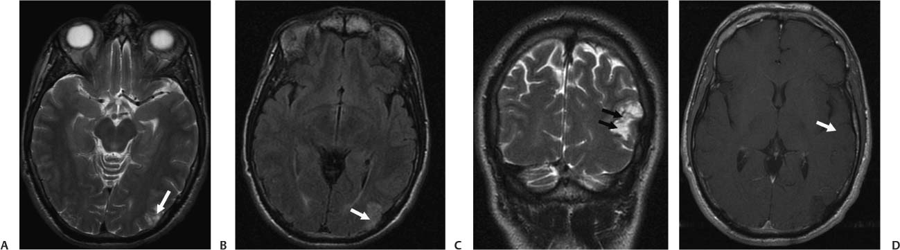
Case 22 A 27-year-old with a history of seizures. (A) Axial T2-weighted image (WI) of the brain shows a lesion in the left parietal lobe (arrow) with a small, cystic, “bubbly” appearance. (B) Axial fluid-attenuated inversion recovery (FLAIR) image demonstrates increased cortical signal in the left parietal lobe (arrow) without surrounding edema. (C) Coronal T2WI demonstrates the cortically based, “bubbly” lesion (arrows) in the left parietal lobe. (D) Axial T1WI with contrast shows no enhancement in the left parietal mass (arrow). • Dysembryoplastic neuroepithelial tumor (DNET):
Clinical Presentation
Imaging Findings
Differential Diagnosis
![]()
Stay updated, free articles. Join our Telegram channel

Full access? Get Clinical Tree


