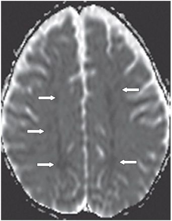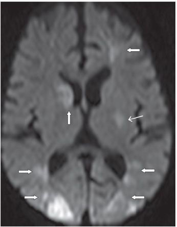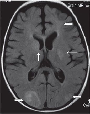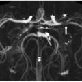


FINDINGS Figures 22-1 and 22-2. Axial DWI with corresponding ADC map through the centrum semiovale. There is almost symmetrical bilateral centrum semiovale irregular linear (rosary-like or string-of-beads pattern) restricted diffusion—hyperintense on DWI with hypointensity on ADC map (arrows). Figures 22-3 and 22-4. Axial DWI with corresponding FLAIR through the basal ganglia. There are bilateral patchy and wedge areas of hyperintensity in bilateral parieto-occipital junctions and around the trigones (posterior arrows), left lentiform nucleus (line arrow), right head of caudate nucleus (right vertical arrow), and left frontal lobe at the anterior cerebral artery (ACA) and middle cerebral artery (MCA) junction (anterior left arrow).
DIFFERENTIAL DIAGNOSIS Watershed infarcts, septic emboli, posterior reversible encephalopathy syndrome (PRES).
DIAGNOSIS Bilateral watershed infarcts.
DISCUSSION
Stay updated, free articles. Join our Telegram channel

Full access? Get Clinical Tree


