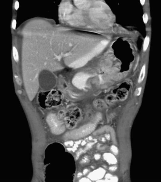
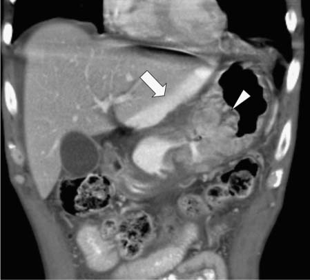
Case 22 A 49-year-old man presents with a history of upper gastrointestinal bleeding and new onset of abdominal pain. Coronal reformatted contrast-enhanced computed tomography image shows a large, expansile mass (arrowhead) within the gastric lumen, adjacent mural thickening, and adjacent extraluminal contrast material (arrow). Free air is present below the middiaphragm. • Gastric adenocarcinoma with perforation: This is the most likely diagnosis, suggested by a fungated, polypoid mass obliterating the gastric lumen and with adjacent mural thickening and extraluminal contrast material. • Lymphoma: This is a possible diagnosis as it may exhibit the same set of possible imaging appearances. • Metastasis to the stomach:
 Clinical Presentation
 Imaging Findings
 Differential Diagnosis
![]()
Stay updated, free articles. Join our Telegram channel

Full access? Get Clinical Tree


