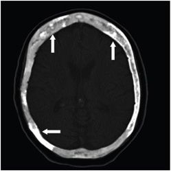
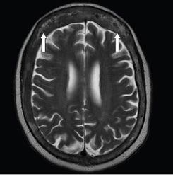
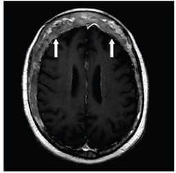
FINDINGS Figure 220-1. Bone scintigraphy. There is abnormal tracer uptake in the calvarium, skull base, and pelvis (arrows). Figure 220-2. NCCT head bone window. There are cortical thickening, abnormal bone matrix, and patchy sclerosis (hyperdensity) (arrows). Figure 220-3. Axial T2WI through the cerebral hemispheres. There are diffuse thickening and heterogeneous marrow intensity of the cranial vault (arrows). Figure 220-4. Axial post-contrast T1WI through the cranial vault. There are marrow replacement, expansion of the diploic space, and patchy enhancement (arrows).
DIFFERENTIAL DIAGNOSIS Paget disease, metastases, fibrous dysplasia.
DIAGNOSIS Paget disease.
DISCUSSION


Stay updated, free articles. Join our Telegram channel

Full access? Get Clinical Tree


