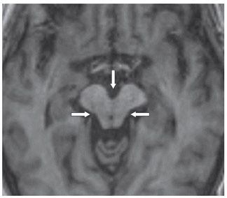
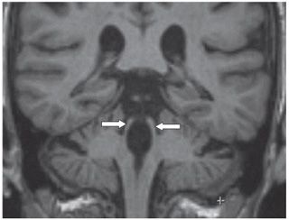
FINDINGS Figure 222-1. Sagittal T1 MPRAGE. There is moderate midbrain/tegmental volume loss (vertical arrow), thinned tectal plate (posterior transverse arrow), mild pontine volume loss (flattening of the basis pontis-chevron) known as “hummingbird sign.” Figure 222-2. Axial MPRAGE through the midbrain. There is widening of the interpeduncular cistern (vertical arrow) and concave appearance of the lateral midbrain (transverse arrows). Figure 222-3. Coronal MPRAGE through the brainstem. There is loss of volume of the superior cerebellar peduncle (arrows).
DIFFERENTIAL DIAGNOSIS


Stay updated, free articles. Join our Telegram channel

Full access? Get Clinical Tree


