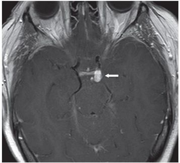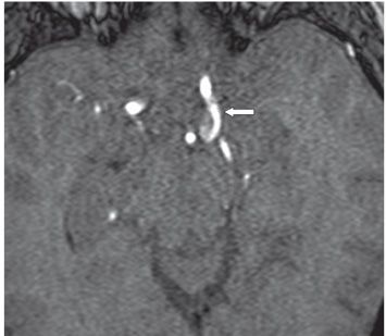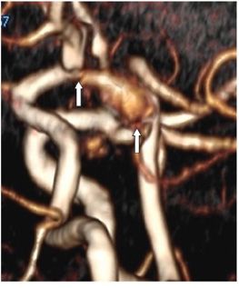


FINDINGS Figure 223-1. Axial MRI FLAIR through the suprasellar cistern. There is diffuse hyperintensity throughout the visualized SAS—suprasellar cisterns (anterior vertical arrows), bilateral sylvian fissures (transverse arrows), and the quadrigeminal/ambient cisterns (posterior vertical arrow) consistent with acute subarachnoid hemorrhage (SAH). A CT scan done earlier on in the day shows basal and convexity SAH and IVH (intraventricular hemorrhage). Figure 223-2. Post-contrast T1WI through the suprasellar cistern. There is an ovoid contrast-enhancing focus measuring 10.4 mm × 6.24 mm on the left in the suprasellar cistern. Figure 223-3. 3D TOF MRA source image through the suprasellar cistern. There is an ovoid outpouch measuring 11.6 mm from the neck to the dome and 3.25 mm at the neck from the left internal carotid artery (ICA) projecting posteriorly between the basilar artery and the left superior cerebellar artery (arrow) consistent with an aneurysm at the level of the posterior communicating artery (P-Com). Figure 223-4. 3D TOF MRA 3D volume rendering. The aneurysm (arrows) is pear shaped and projects posteriorly from the left ICA.
DIFFERENTIAL DIAGNOSIS ICA anterior choroidal artery aneurysm, posterior communicating artery aneurysm (P-Com-A).
DIAGNOSIS P-Com-A.
DISCUSSION
Stay updated, free articles. Join our Telegram channel

Full access? Get Clinical Tree


