

FINDINGS Case previously published BJR 2006; 79: e140-e144.
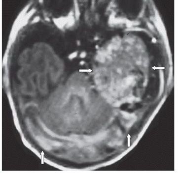
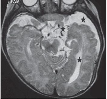
Figure 226-1. Axial FLAIR through the midbrain. There is a large collection of tubular signal voids measuring about 5 cm in maximum dimension in the left temporal lobe (transverse arrows). There is mild mass effect on the midbrain. This tangle of blood vessels drains into the large torcula (vertical arrow). There is hyperintense brain parenchyma interspersed within the mass presumably gliosis. Figure 226-2. Axial T1WI through the temporal lobes. There is a large, ovoid, and heterogeneous left temporal lobe well-defined mass (transverse arrows). The bilateral transverse sinuses are large (vertical arrows). Figures 226-3
Stay updated, free articles. Join our Telegram channel

Full access? Get Clinical Tree


