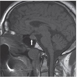
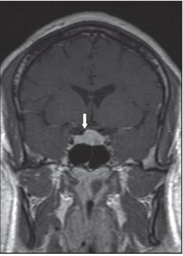
FINDINGS Figure 228-1. Sagittal MR post-contrast T1WI. There is diffusely enhancing infiltrative lesion in the pituitary gland, stalk, and suprasellar region (arrow). Figure 228-2. Sagittal non-contrast T1WI in a companion case. There is an enlarged pituitary gland (arrow). Figure 228-3. Coronal post-contrast T1WI in the companion case. There is homogeneous enhancement of the enlarged pituitary gland (arrow).
DIFFERENTIAL DIAGNOSIS Metastases, meningitis, meningioma, tuberculosis, lymphoma, leukemia, sarcoidosis, and histiocytosis. When the brain is affected, multiple sclerosis, Lyme disease, and vasculitis should be considered.
DIAGNOSIS Pituitary axis neurosarcoidosis.
DISCUSSION


Stay updated, free articles. Join our Telegram channel

Full access? Get Clinical Tree


