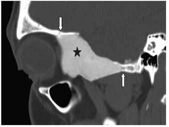
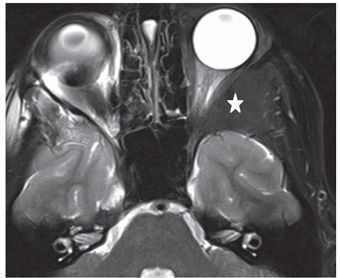
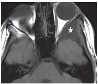
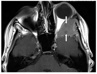
FINDINGS Figures 229-1 and 229-2. Axial and reformatted sagittal NCCT through the orbits. There is expansion and sclerotic (ground-glass) appearance of the left greater wing of sphenoid bone (stars). There is abrupt sharp transition between the lesion and surrounding bone (arrows). There is mild encroachment on the left orbit with mild proptosis. Figures 229-3 and 229-4. Axial T2WI and T1WI, respectively, through the lesion. There is homogeneous hypointensity of expanded thickened left greater wing of sphenoid bone (stars). There is compression of the left lateral rectus and narrowing of the left orbital apex. Figure 229-5. Axial post-contrast T1WI through the lesion. There is almost homogeneous contrast enhancement of the lesion with two tiny poorly enhancing foci (arrows).
DIFFERENTIAL DIAGNOSIS Osteoblastic metastasis, fibrous dysplasia (FD), Paget disease, intraosseous meningioma.
DIAGNOSIS Fibrous dysplasia (FD).


Stay updated, free articles. Join our Telegram channel

Full access? Get Clinical Tree


