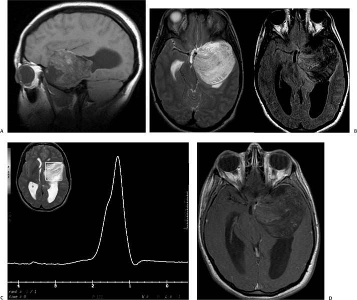
Case 23 A 17-year-old girl with a history of seizures. (A) Sagittal T1-weighted image (WI) shows a mass in the anterior aspect of the temporal fossa with areas of T1 hyperintensity (arrow). The mass is compressing the adjacent parenchyma. (B) Axial T2-weighted and fluid-attenuated inversion recovery (FLAIR) images demonstrate heterogeneous signal in the left temporal fossa mass. The mass shows areas of increased signal on the FLAIR image (arrow). The location of the lesion is extra-axial. It compresses the temporal lobe and cerebellar peduncle (arrowheads), displacing vessels without encasing them. (C) Magnetic resonance (MR) spectroscopy shows a high lipid peak with a broad base from –0.9 to –1.3 parts per million (ppm). (D) Axial T1WI with contrast shows no enhancement of the mass; the hyperintense regions (arrow) were bright in the nonenhanced sequences (related to fat). • Nonruptured dermoid cyst:
Clinical Presentation
Imaging Findings
Differential Diagnosis
![]()
Stay updated, free articles. Join our Telegram channel

Full access? Get Clinical Tree


