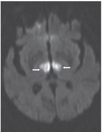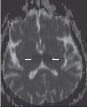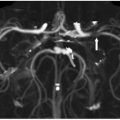

FINDINGS Figure 23-1. Axial FLAIR image through the thalami. There is bilateral almost symmetrical medial thalamic hyperintensity (arrows). Figures 23-2 and 23-3. Corresponding axial DWI and ADC map, respectively, through the thalami. The lesions restrict diffusion—hyperintense on DWI with low ADC—consistent with acute infarctions.
DIFFERENTIAL DIAGNOSIS Basilar artery thrombosis, deep cerebral venous sinus thrombosis, artery of Percheron infarctions.
DIAGNOSIS Bilateral paramedian thalamic infarcts from occlusion of the artery of Percheron.
DISCUSSION Acute arterial infarctions of both inferior and medial thalami most often result from occlusion of the rostral basilar artery, which also commonly affect the midbrain and portions of the temporal and occipital lobes (supplied by the posterior cerebral artery) or the cerebellum (supplied by branches of the vertebrobasilar arterial system).
One of the rare anatomical variants of the posterior circulation is the artery of Percheron (Table 38-1
Stay updated, free articles. Join our Telegram channel

Full access? Get Clinical Tree


