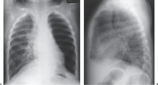
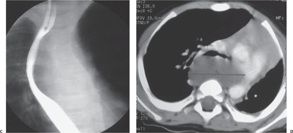
Case 23 An 8-year-old boy with chest pain and difficulty swallowing. (A,B) Frontal chest radiograph demonstrates right paratracheal opacity. The left lung is hyperlucent. Lateral chest radiograph demonstrates a large mass posterior to the trachea (arrow), associated with tracheal narrowing. (C) Esophagogram demonstrates that the esophagus is displaced to the right by an extramural mass (arrow). (D) Axial post-contrast computed tomography (CT) image shows a large, low-density mass at the level of the carina. It narrows the left main bronchus (arrow). The right lung is now hyper-expanded. • Bronchogenic cyst:
Clinical Presentation
Further Work-up
Imaging Findings
Differential Diagnosis
![]()
Stay updated, free articles. Join our Telegram channel

Full access? Get Clinical Tree


