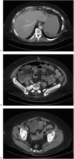
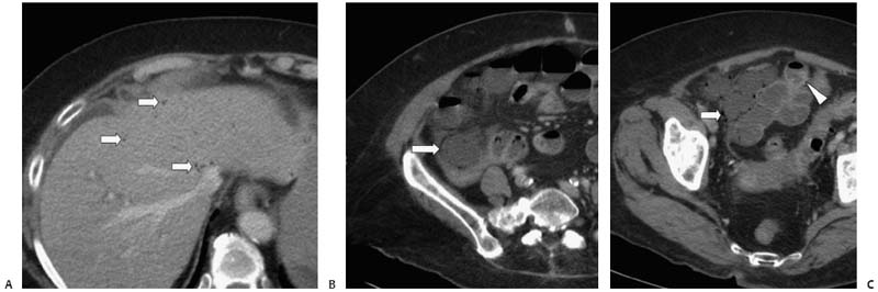
Case 23 A 61-year-old woman presents with acute abdominal pain and fever. (A) Contrast-enhanced abdominal computed tomography (CT) image shows multiple punctate, hypodense foci (arrows) in the liver that are difficult to characterize, given their small size, but are suspicious for portal venous gas. (B) Pelvic image shows fluid-filled cecum with pneumatosis (arrow) and mesenteric fat stranding. (C) More caudal image shows multiple loops of distended terminal ileum, some with pneumatosis (arrow) and wall thickening (arrowhead). • Acute mesenteric ischemia (AMI): This is the most likely diagnosis, given the findings of portal venous gas, pneumatosis, mesenteric fat stranding, and thickened, distended, fluid-filled terminal ileum and cecum. • Infection of the ileum and cecum ( Yersinia infection, tuberculosis, actinomycosis, and amebiasis):
 Clinical Presentation
 Imaging Findings
 Differential Diagnosis
![]()
Stay updated, free articles. Join our Telegram channel

Full access? Get Clinical Tree


