The Thalamoperforating Arteries
23.1 Case Description
23.1.1 Clinical Presentation
A 48-year-old patient presented with acute onset of confusion and ataxia to the emergency room.
23.1.2 Radiologic Studies
See ▶ Fig. 23.1, ▶ Fig. 23.2.
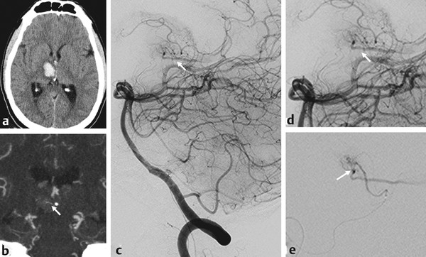
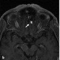
Fig. 23.1 Unenhanced CT (a) demonstrated a right thalamic hemorrhage. As there was no evidence for microangiopathy on the CT and no history regarding cardiovascular risk factors, CTA (b) and, subsequently, digital subtraction angiography were performed. In retrospect, the CTA demonstrated a tiny asymmetry in the vascularity of the thalamus showing a vessel coursing through the area of hemorrhage (arrow). Vertebral artery injection (b,c) (lateral view with magnification of the area of interest in d) demonstrated a microshunt that was only visible because of early venous shunting (arrows in b,c,d). Superselective catheterization of the principal group of the inferolateral thalamoperforators arising from the P2 segment of the PCA (e) demonstrated the shunt (arrow). After more distal catheterization, the shunt was treated with liquid embolic agent embolization. Case continued in ▶ Fig. 23.2.
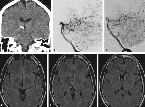
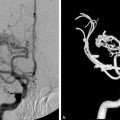
Fig. 23.2 After the procedure, the patient awoke without new neurological deficits. Coronal unenhanced CT (a) demonstrates the glue cast and angiographic controls immediately after the procedure (vertebral artery lateral view) (b) and, after 1 year (c), demonstrated complete obliteration of the shunt. Axial fluid-attenuated inversion recovery weighted scans (d,e,f) in adjacent cuts after embolization demonstrate the sequelae of the hemorrhage and no focal ischemia.
23.1.3 Diagnosis
Thalamic micro-arteriovenous malformation (AVM) fed by the principal group of the inferolateral thalamoperforators.
23.2 Anatomy
There are three major thalamoperforating artery (ThPA) groups that supply the different thalamic vascular territories. The nomenclature varies, but for this chapter they are referred to as the tuberothalamic, paramedian, and inferolateral arteries. These groups have also been referred to as the anterior ThPA, posterior ThPA, and thalamogeniculate arteries. There is high individual variability in the number, origin, and arterial territory supplied by each group. The small size of the perforator branches makes them often difficult to visualize, but they can be seen on lateral angiograms.
The tuberothalamic arteries arise from the middle third of the posterior communicating artery (PcomA) and supply the ventral section of the thalamus, which includes the reticular nucleus, the ventral anterior nucleus, the rostral section of the ventrolateral nucleus, the ventral pole of the medial dorsal nucleus, the mamillothalamic tract, the ventral amygdalofugal pathway, the ventral section of the internal medullary lamina, and the anterior thalamic nuclei. They can be divided into interpeduncular, hypothalamic, and thalamic segments. The interpeduncular segment loops posterior to the optic tract. It is extremely short and sometimes nonexistent. The hypothalamic segment enters the hypothalamus in close relationship to the lateral wall of the third ventricle. The thalamic segment appears as a characteristic cluster of vessels that supplies the nuclei mentioned earlier. The tuberothalamic group can be absent in ~30% (up to 60%, according to some series) of the normal population. In these cases, the tuberothalamic arterial territory is taken over by the paramedian group.
The paramedian arteries are usually two arteries that arise from the P1 segment of the posterior cerebral artery (PCA), but their number can range between one and five. They supply the dorsomedial nucleus, internal medullary lamina, and intralaminar nuclei and are presumed to represent the most cranial group of paramedian arteries of the basilar artery. They can be divided into interpeduncular, mesencephalic, and thalamic segments. The interpeduncular segment has a characteristic short but very sinuous course. The mesencephalic segment, in contrast, has a characteristically straight course as it traverses the midbrain, allowing the differentiation between the segments. The thalamic segment appears as a cluster of vessels, similar to the one from the tuberothalamic arteries. The paramedian arteries can arise as a pair from each P1, but also as a common trunk, thus supplying the thalamus bilaterally (artery of Percheron) (▶ Fig. 23.3). When the tuberothalamic arteries are absent, the paramedian arteries take over their territory.
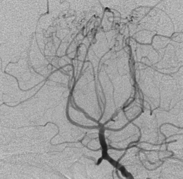
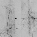
Fig. 23.3 A 52-year-old man was evaluated for moyamoya disease. Anteroposterior (AP) left vertebral artery angiogram revealed a single right arterial trunk supplying the paramedian thalami bilaterally, in keeping with an artery of Percheron. The patient showed an unusually straight course of the interpeduncular segment.
The inferolateral arteries are between 5 and 10 arteries that arise from the P2 segment of the PCA. They are subdivided into three groups: medial geniculate, principal inferolateral, and inferolateral pulvinar. The medial group supplies the external half of the medial geniculate nucleus. The principal inferolateral group penetrates between the geniculate bodies, ascends in the lateral medullary lamina, and supplies the major part of the ventral posterior nuclei and parts of the ventrolateral nucleus. The inferolateral pulvinar group supplies the rostral and lateral parts of the pulvinar and the lateral dorsal nucleus.
Stay updated, free articles. Join our Telegram channel

Full access? Get Clinical Tree


