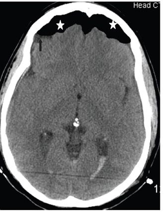
FINDINGS Figures 230-1 and 230-2. Axial contiguous NCCT through the inferior frontal lobes. There are bilateral frontal extraaxial hypodensities (air collections) (stars) with compression of the frontal lobes. The frontal lobes “peak” into the air collections with a midline separation of the frontal lobes (white arrows). There is air–fluid level laterally in the collections (black arrows). Intraventricular hemorrhage due to diffuse axonal injury (DAI) is present in the trigones in Figure 230-2.
DIFFERENTIAL DIAGNOSIS N/A.
DIAGNOSIS Pneumocephalus (tension pneumocephalus).
DISCUSSION


Stay updated, free articles. Join our Telegram channel

Full access? Get Clinical Tree


