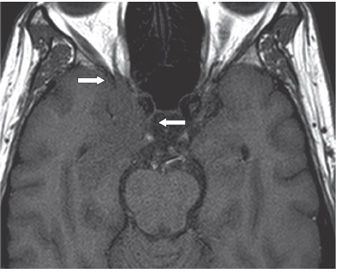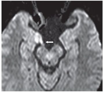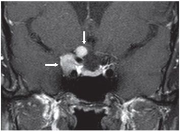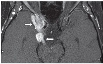


FINDINGS Figure 231-1. Coronal T2WI through the chiasm. There is enlargement and mild hyperintensity of the optic chiasm on the right (vertical arrow) which extends on the other images (not shown) along the right optic nerve into the optic foramen. There is hypointense thickening of the right cavernous sinus (transverse arrow). Figure 231-2. Axial T1WI through the suprasellar cistern. There is hypointensity of the right uncus extending into the right superior orbital fissure (arrows). Figure 231-3. Axial DWI through the suprasellar cistern. There is mild restricted diffusion in the nodular right third cranial nerve (arrow). The mass compresses the right uncus laterally. Figure 231-4. Coronal post-contrast T1WI through the anterior sella turcica. There is avid nodular contrast enhancement of the right optic nerve (vertical arrow) which on the other images (not shown) extends posteriorly to the chiasm and anteriorly to the right orbital apex. There is also avid contrast enhancement of the enlarged right cavernous sinus (transverse arrow). Figure 231-5
Stay updated, free articles. Join our Telegram channel

Full access? Get Clinical Tree


