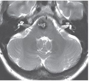
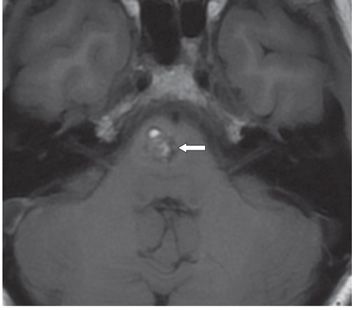
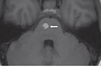
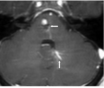
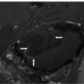
FINDINGS Figure 232-1. Axial GRE through the inferior pons. There is a 1.7-cm minimally irregular mass slightly to the right in the basis pontis (arrow). Mass has a very thick dark (blooming hemosiderin) rim with a heterogeneous core. Figure 232-2. Axial T2WI through the mass. The irregular dark rim is thinner with the heterogeneous core slightly wider compared with the GRE. There is no surrounding edema or mass effect. Figures 232-3 and 232-4. Axial non-contrast T1WI through the mass at two contiguous levels. There are two components to the mass. There is a heterogeneous component (popcorn pattern) inferiorly and posteriorly (arrow in Figure 232-3) and a hyperintense component (subacute hemorrhage) superiorly and anteriorly (arrow in Figure 232-4), both with a thin hypointense rim. Figure 232-5. Axial post-contrast T1WI through the mass. There is a thin peripheral rim enhancement posteriorly (transverse arrow) associated with a contrast-enhancing DVA in the left cerebellum (vertical arrow).
DIFFERENTIAL DIAGNOSIS Hematoma, cerebral cavernous malformation (CCM), developmental venous anomaly (DVA), hemorrhagic tumor.


Stay updated, free articles. Join our Telegram channel

Full access? Get Clinical Tree


