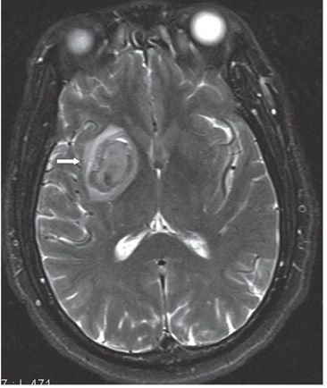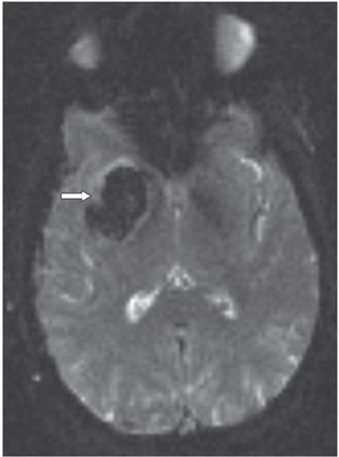

FINDINGS Figure 233-1. Axial NCCT through the basal ganglia. There is an ovoid right lentiform nucleus/external capsular well-defined homogeneous hyperdensity (arrow) without significant mass effect on adjacent structures or midline shift. Findings consistent with acute hematoma. Figure 233-2. Axial T2WI through the basal ganglia. There is an ovoid lamellated heterogeneous mass in the right lentiform nucleus/external capsule (arrow). The outside ring of hyperintensity is due to perilesional edema. Figure 233-3. Axial GRE through the basal ganglia. There is a homogeneous hypointense mass in the right lentiform nucleus/external capsule (arrow). There is a surrounding thin rim of hyperintensity (edema).
DIFFERENTIAL DIAGNOSIS Hematoma due to vascular malformations, deep venous thrombosis, hemorrhagic tumors including metastasis, amyloid angiopathy, anticoagulation, and drug abuse (typically methamphetamine and cocaine).
DIAGNOSIS Hypertensive basal ganglia hematoma (HBGH).
DISCUSSION
Stay updated, free articles. Join our Telegram channel

Full access? Get Clinical Tree


