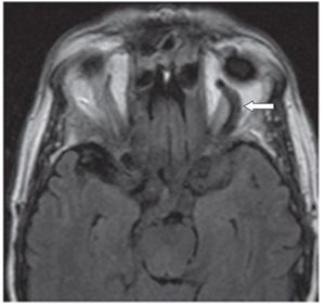
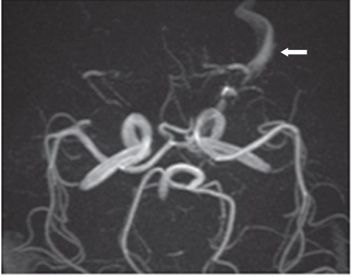
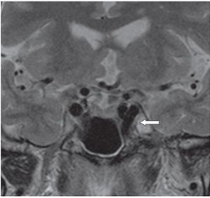
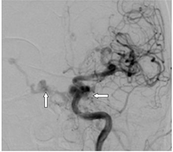
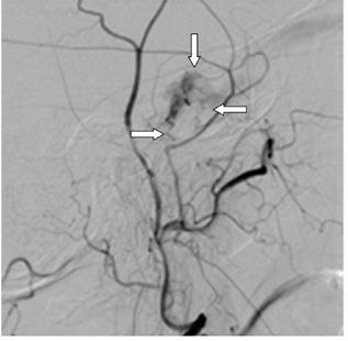
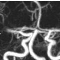
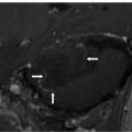
FINDINGS Figure 234-1. Axial NCCT through the cavernous sinuses. There is a subtle outward bulge of the left cavernous sinus (arrow). Figure 234-2. Axial FLAIR through the orbits. There is an enlarged and tortuous left superior ophthalmic vein (arrow). Figure 234-3. 3D TOF MRA. There is visualization of an enlarged left superior ophthalmic vein (arrow) similar to the arterial structures. Figure 234-4. Coronal T2WI shows an additional “flow void” in the left cavernous sinus (arrow). Figure 234-5. Anteroposterior DSA. There is early opacification of the cavernous sinuses (arrows) during a selective injection of the left internal carotid artery (ICA). Figure 234-6. Lateral DSA of selective left external carotid artery (ECA). There is ECA feeders from the left middle meningeal artery and sphenopalatine artery (arrows).
DIFFERENTIAL DIAGNOSIS Tolosa-Hunt syndrome, cavernous sinus thrombosis, carotid cavernous sinus fistula, thyroid ophthalmopathy, pseudotumor or retrobulbar masses (such as metastases).
DIAGNOSIS Carotid-cavernous sinus fistula (CCF).
DISCUSSION


Stay updated, free articles. Join our Telegram channel

Full access? Get Clinical Tree


