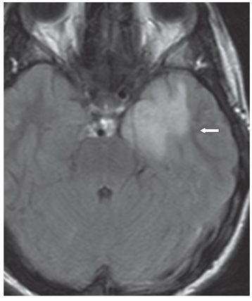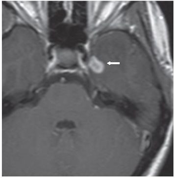

FINDINGS Figure 235-1. Axial post-contrast CT through the temporal lobes. There is a vague left temporal lobe hypodensity with a possible vague ring enhancement in the left parasellar region (arrow). Figure 235-2. Axial FLAIR MR through the temporal lobes. There is a confluent medial left temporal lobe hyperintensity with lateral peripheral finger-like projections consistent with vasogenic edema (arrow). Figure 235-3. Axial post-contrast T1WI through the temporal lobes. There is a 1.5-cm thick ring enhancement in the medial left temporal lobe just lateral to the left cavernous sinus (arrow). MRA obtained subsequently showed no aneurysm.
DIFFERENTIAL DIAGNOSIS Ganglioglioma, high-grade astrocytoma, abscess.
DIAGNOSIS Langerhans cell histiocytosis (LCH).
DISCUSSION
Stay updated, free articles. Join our Telegram channel

Full access? Get Clinical Tree


