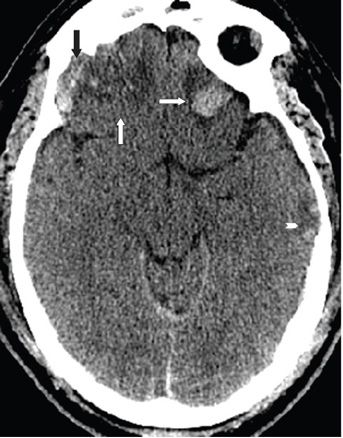
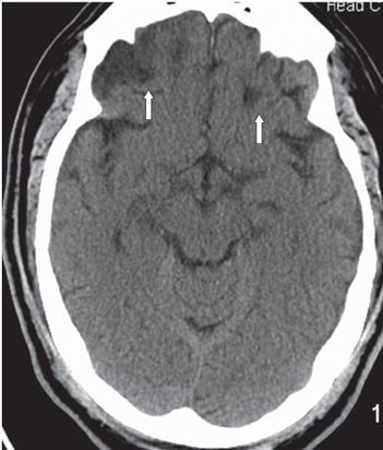
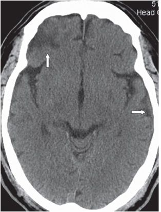
FINDINGS Figures 236-1 and 236-2. Axial contiguous NCCT through the inferior frontal lobes. There is a right frontal lobe hypodensity just above the right orbital roof (vertical arrows) consistent with contusion. There is a small lateral irregular heterogeneous area consistent with the “pepper-and-salt” pattern of hemorrhagic contusion (black arrows). In the left frontal lobe, there is a round hyperdensity surrounded by a hypodense halo above the left orbital roof (transverse arrows) consistent with hematoma. There is a smaller mixed density cortical lesion in the left temporal lobe (chevrons) consistent with hemorrhagic contusion. Figures 236-3 and 236-4. Contiguous NCCT through the inferior frontal lobes 6 months after Figures 236-1 and 236-2. There is bifrontal and left temporal sharply demarcated hypodensities in areas of previous contusions and hematoma (arrows) consistent with encephalomalacia. There is generalized widening of the subarachnoid spaces indicating volume loss.
DIFFERENTIAL DIAGNOSIS Contusions, infarcts.
DIAGNOSIS Multifocal hemorrhagic brain contusions.


Stay updated, free articles. Join our Telegram channel

Full access? Get Clinical Tree


