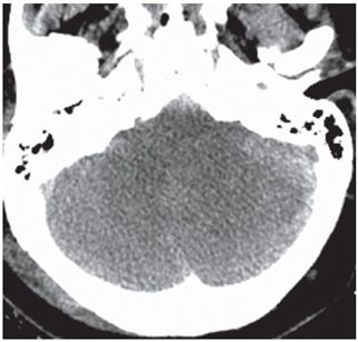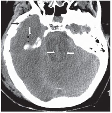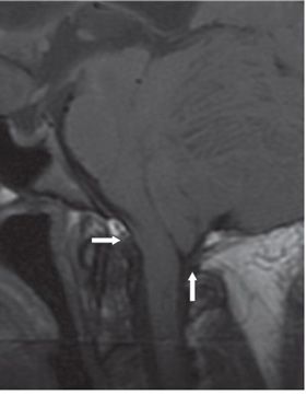


FINDINGS Figure 238-1. Axial NCCT through the foramen magnum. There is complete effacement of the cisterna magna and the cerebrospinal fluid (CSF) space surrounding the upper spinal cord (arrow). Figure 238-2. Axial NCCT through the inferior posterior fossa. There is complete obliteration of the posterior fossa CSF spaces including the fourth ventricle, the cerebellopontine angle (CPA) cisterns, and the pericerebellar spaces by the swollen brainstem and cerebellum. Figure 238-3. Axial NCCT through the upper pons. There is swelling and mild anteroposterior elongation of the pons. There is swelling of the cerebellum and visualized supratentorial brain with obliteration of all CSF spaces. There are hemorrhages in the pons (transverse white arrows) and around the tentorium. In the right temporal lobe, pneumocephalus is present (vertical white arrow). There is also a right temporal extraaxial acute hemorrhage and pneumocephalus (black arrow). A transtentorial herniation is present in this patient (not shown). Figure 238-4
Stay updated, free articles. Join our Telegram channel

Full access? Get Clinical Tree


